Biomechanics of Tissue Adds Essential Information to Optical Imaging
A family of techniques combining optical examination of biological tissues with measurements of their mechanical properties is set to assist with a number of current medical challenges, including recovery from heart attacks and treatment of corneal disease.
The potential impact of these technologies was recognized at SPIE Photonics West in February by the holding of the seventh Optical Elastography and Tissue Biomechanics conference, co-chaired by Kirill Larin of the University of Houston's Department of Biomedical Engineering, and Giuliano Scarcelli from the University of Maryland.
"This conference was inaugurated in 2014, at a time when the field of biomechanics was starting to grow and research into combining biomechanical measurements with optical imaging was expanding," said Larin.
"Today there is an established body of published research and regular breakthroughs in this field, as people start to recognize that biomechanics can reveal more information about tissues than optical examination alone, and bring additional contrast to images obtained by optical techniques."
The motivation behind this growth is the increased understanding of ways in which biomechanical properties, such as tissue stiffness, can play a significant role in both the development of disease and subsequent recovery from treatment. Those properties could also ultimately be a predictive marker for a number of diseases and degenerative conditions, including cancer.
One example would be the ways in which myocardial infarctions - heart attacks - leave the damaged areas of the heart as scarred tissue, with distinctly different biomechanical properties from those of healthy cells. Restoring the original attributes and healing the tissues is one route to treatment and therapy, but ways to accurately assess the tissue mechanics are an essential part of the process.
"The only way we can monitor the success of such therapies is by measuring the mechanical properties and seeing how they change as the heart heals," commented Larin.
Additional contrast
The technique being brought to bear on myocardial infarction is optical coherence elastography (OCE), in which optical coherence tomography (OCT) is used to image tissue while it is under some form of transient local mechanical deformation and subsequent recovery. This exploits the basic difference between image contrast created by mechanical action, and intrinsic optical contrast: an OCT scan assesses differences in scattering from a target area, while mechanical contrast requires only scattering above a basic OCT threshold, and without it necessarily varying over a sample.
"OCT plus some additional contrast mechanism is an inherently powerful approach, and one beauty of OCE is that OCT is already well established in clinics," said Larin. "OCE can fill a gap in non-destructive imaging techniques, combining field-of-view in the millimeter or centimeter range with spatial resolution from microns to millimeters."
The applied external force needed in OCE can be delivered by different excitation methods, both contact and non-contact. Each has its advantages, and Kirill Larin's lab at Houston is working on non-contact and completely non-invasive stimulation techniques, which are likely to be best suited for eventual translation into clinics and reducing patient discomfort.
Given OCT's existing wide acceptance in ophthalmology, an OCE platform using a non-contact excitation source could be a versatile way to assess the properties of the cornea in cases of keratoconus, where the cornea has become deformed, and to monitor the corneal cross-linking treatments used to address the disease.

OCE combines optical and mechanical data to assist with diagnoses. Credit: Zvietcovich, F., Pongchalee, P., Meemon, P. et al, "Reverberant 3D optical coherence elastography maps the elasticity of individual corneal layers," Nat Commun 10, 4895 (2019).
It may even be possible to assess corneal biomechanics without any external stimulation at all, using instead the tiny physical changes caused by the natural heartbeat of a patient, and the pulsed blood flow in the cornea it creates. Other techniques showing promise include shear wave elastography (SWE), the topic of an invited keynote during the BIOS conference from Mathias Fink of France's Langevin Institute.
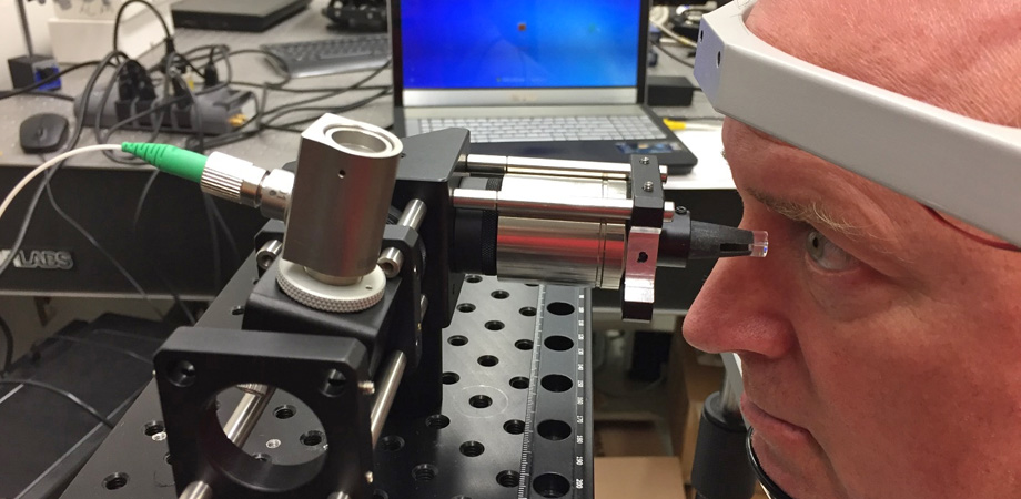
SWE uses external stimulation to create shear waves propagating through the tissue, and an ultrasound pulse has been the traditional way to create these waves. Larin's project group aims to develop alternative methods, and learn from the more established ultrasound approach. "SWE measures mechanical waves produced on the tissue surface or inside the tissue, and allows you to see straightforward relationships between different properties," he said. "In my lab we use a tiny puff of air to produce shear waves of sub-micron amplitude in the cornea, so small that patients do not feel it; but OCT is sensitive enough to detect and image the contrast that those waves produce."
Also on the BIOS agenda is Brillouin elastography, a label- and contact-free method which exploits inelastic scattering of light by acoustic waves in a medium, when those waves are induced by thermal or other excitation. Current areas of research in this technique include ways to address the inherent weakness of the measured scattered signal, which can lead to long data acquisition times and photodamage to the tissues.
Some profitable synergies are expected to develop between these different biomechanical techniques, according to Larin. A combination of OCE and Brillouin spectroscopy could potentially be used to measure the physical properties of the lens of the eye, by incorporating the quantitative nature of OCE with the ability of Brillouin spectroscopy to produce 3D data from deeper layers of target tissues. Directing focused acoustic radiation to the lens of the eye could allow quantitative elastography of the lens in 3D, assisting studies of ubiquitous conditions such as presbyopia.
Niche applications
All these techniques are now gearing up for clinical translation, a process expected to be relatively smooth for a method such as OCE, where the underlying optical instrumentation is already tried and tested. "Each technique has different applications and will take a slightly different route to market," said Kirill Larin.
"Most clinics already employ OCT, for example, and simple modifications can enable the use of OCE and imaging of mechanical contrast, rather than just structural images."
Although no commercial platforms based on tissue biomechanics have come to market as yet, the translation process will soon be bearing fruit. Larin currently has a clinical system being trialed in a hospital, while studies on keratoconus patients are bringing the techniques into the ophthalmology space.
"This field is evolving fast, and with the patents now in place commercialization is very close," he commented. "I am sure that in five years these biomechanical platforms will be in commercial use. With so many potential applications, it is only a matter of time."
Tim Hayes is a freelance writer based in the UK. He was previously industry editor of optics.org and Optics & Laser Europe magazine. A version of this article appeared in the 2020 Photonics West Show Daily.
Related SPIE content:
Microscale imaging of breast tumor margins using optical coherence elastography
David Sampson: Optical coherence elastography may increase efficiency of cancer surgery
Structure imaging of biological tissue by optical coherence elastography
Longitudinal elastic wave imaging using nanobomb optical coherence elastography
Correlation of optical coherence elastography with clinical evaluation of systemic sclerosis
| Enjoy this article? Get similar news in your inbox |
|


