Optipedia • SPIE Press books opened for your reference.
Explanation of cornea from Field Guide to Visual and Ophthalmic Optics
Excerpt from Field Guide to Visual and Ophthalmic Optics
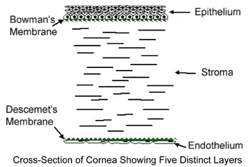
Epithelium
Thin surface layer of cells 50–100 µm thick on the front of the cornea that blocks foreign bodies from entering the cornea and absorbs oxygen and nutrients for the underlying layers. These cells regenerate quickly if they are damaged by trauma or surgery.
Bowman’s Membrane
Collagen boundary roughly 12 µm thick that divides the epithelium and the underlying stroma.
Stroma
Internal material of the cornea composed mainly of cross-linked collagen bands. The ordering of these bands is somewhat regular to promote transparency. The thickness of the stroma is about 500 µm. The collagen fibers are somewhat regularly organized, introducing corneal birefringence.
Descemet’s Membrane
Collagen boundary roughly 4 to 10 µm thick that separates the endothelium and the stroma.
Endothelium
Thin surface layer of cells 5 µm thick on the back of the cornea that regulates corneal nutrition and removes excess water from the cornea to maintain its clarity. Unlike the epithelial cells, these cells do not regenerate.
The cornea does not have a direct blood supply, so it must exchange its nutrients and waste products through its front and back surfaces. Damaging or interfering with these transfer mechanisms can lead to corneal edema (swelling) and opacities.
J. Schwiegerling, Field Guide to Visual and Ophthalmic Optics, SPIE Press, Bellingham, WA (2004).
View SPIE terms of use.
