Predicting Cancer’s Return: Pump-probe analysis reveals metastatic potential
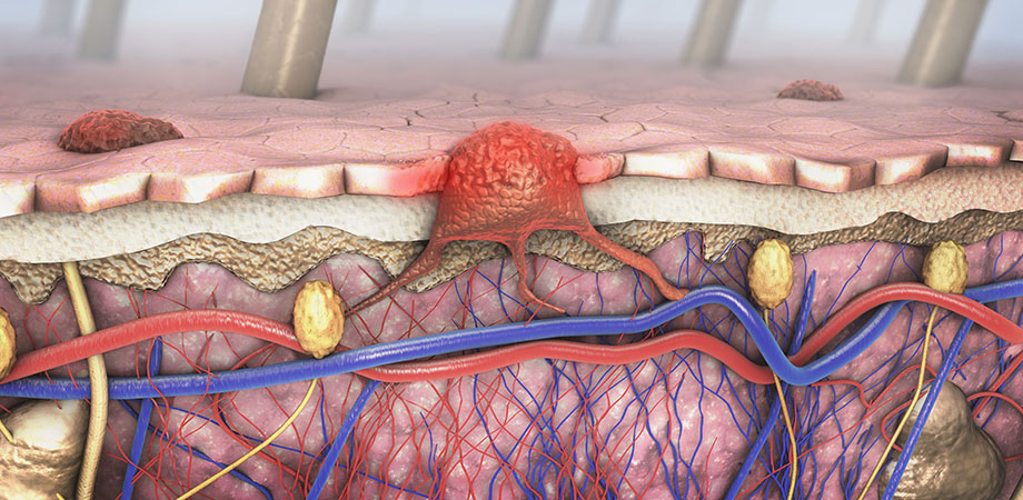
Skin cancer is probably one of the easiest cancers to treat: the cancer is visible on the skin, and it can usually be removed with a simple operation. However, malignant melanoma should not be taken lightly. In the US, about 96,000 people per year are diagnosed with melanoma, and 7,000 die from the disease. In sunnier climes, like Australia, the rates are two to three times higher.
For those diagnosed early, a stage I melanoma has a high five-year survival rate, but risk remains. It is very hard to predict which melanomas are going to be metastatic. As a result, more people die from metastatic melanoma long after a stage I diagnosis than those who are initially diagnosed at stage IV. A tool that could more accurately predict the chance of reoccurrence and metastasis at stage I would be very useful indeed.
Within the last decade, researchers have discovered that, for some reason, melanoma cells respond very differently to laser light than normal cells and certain features were correlated with the risk of metastasis. The underlying reason for that difference is explained in a recently published paper by Kuk-Youn Ju and colleagues from Duke University.
Pumping up the melanin
Their work is based around a technique called pump-probe spectroscopy. Pump-probe spectroscopy explores the dynamics of molecular systems. A laser pulse, called the pump, excites a sample. Over time, the sample will relax back to its ground state, and the goal of the pump probe spectroscopist-including this research team-is to understand the path that the sample takes back to the ground state. This is done by hitting the sample with a second laser pulse, called the probe, which arrives slightly later than the pump pulse. The absorption of the probe pulse reveals information about the sample state at that moment in time.
The sample state can be obtained by repeating the experiment with varying time differences between the pump and probe, as well as different pump or probe wavelengths. This multidimensional spectroscopy reveals the rich dynamics of energy absorption, transfer, and dissipation in molecular systems.
To understand why melanoma cells have a different pump-probe response than normal skin cells, the researchers focused on the main source of pigment in a melanoma cell: eumelanin. Eumelanin, which also exists in healthy skin cells, is a planar molecule that, under normal circumstances, stacks into layers to form large (50-300 nm) blobs. However, the stacking matters: a disordered aggregate will absorb and dissipate energy differently compared to an ordered stack, which should be observable in pump-probe spectroscopy. On the diagnosis side, the disintegration of order may indicate the likelihood of metastasis.
To test this idea, the researchers compared different eumelanin preparations containing either ordered stacks ("thick oligomer stacks") or disordered stacks ("thin oligomer stacks"). The team's pump-probe experiments showed that ordered stacks of eumelanin were subject to ground state bleaching. This means that the vast majority of the sample is excited by the pump, making it more transparent to the subsequent probe. On the other hand, the disordered eumelanin samples showed evidence of excited-state absorption. This means that after the pump has driven the eumelanin into an excited state, the probe is also absorbed, further exciting the eumelanin.
This effect, the researchers showed, is due purely to the way the eumelanin assembles. Ordered structures can quickly spread the energy delivered by the pump into other states-most likely into vibrational modes-emptying the excited state and reducing probe absorption. Disordered stacks are slower to lose their energy, leaving the eumelanin in an excited state and available to be further excited by the incoming probe pulse.

Proposed model for the chemical heterogeneity of eumelanin in the progression of melanoma and its relation to the pump-probe signal. Credit: doi.org/10.1117/1.JBO.24.5.051414
Disordered eumelanin predicts cancer's return
In this study, excited-state absorption dominant signals are shown to be originated from disordered stacks and fragments of eumelanin. Furthermore, pump-probe spectroscopy on biopsies shows that the change from order to disorder is strongly linked to the probability of someone getting melanoma in the future.
This research provides an important link between spectroscopic observation and changes to the biochemistry of the cell. It may give surgeons and doctors the justification they need to more aggressively treat the initial melanoma and to justify a long-term observation program to catch reoccurrence earlier. It may also be worth future investigations to determine if early signs of eumelanin degradation are a predictor for future melanoma. If they are, then we would have a powerful diagnostic tool: pump-probe spectroscopy analysis would help predict both the first occurrence of melanoma and the probability of metastatic melanoma returning in later years.
Read the original article in the Journal of Biomedical Optics, doi.org/10.1117/1.JBO.24.5.051414
Chris Lee is a physicist and writer living and working in Eindoven, the Netherlands.
| Enjoy this article? Get similar news in your inbox |
|


