Taking the Pulse of the Eyes: OCT Measures the Mechanics of Eyes in Response to Heartbeats
Life is measured in heartbeats, so they say—and heartbeats may soon help measure the health of our eyes. If doctors can measure pulse in the eye to diagnose corneal pathologies, the results may save vision and may also save lives.
Every heartbeat sends a pulse wave of blood, inducing very small yet measurable changes in the eye volume, with a resultant momentary increase in intraocular pressure. Working from the postulate that changes in the elasticity or stiffness of the eye may signal significant structural or functional changes, researchers are seeking ways to accurately measure ocular biomechanics.
According to SPIE fellow Dr. Kirill Larin, Cullen College of Engineering Professor of Biomedical Engineering at the University of Houston, "Accurate measurement of corneal biomechanics would not only influence our clinical interpretation of diagnostic tests, for example by measuring intraocular pressure or assessing effects of drug therapies, but also predict the onset of posterior eye diseases, such as glaucoma." Currently, there is no available reliable method to perform quantitative measurement of corneal elasticity in living eyes.
To meet this clinical need, Dr. Larin's group is developing a new approach for completely "no-touch" assessment of corneal biomechanics. Convenient for the patients, this approach would advance medical understanding of corneal disorders, allow developing novel clinical therapies and interventions, and improve the outcome of current surgical and therapeutic interventions. The latest step in their research is published in SPIE's Journal of Biomedical Optics (JBO).
Stretchy eyes
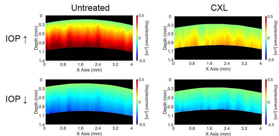
In 2013, Rainer A. Leitgeb first demonstrated the possibility of measuring the motion of ocular tissue due to the heartbeat using phase-sensitive optical coherence tomography (OCT). Inspired by that work, Kirill Larin's group is currently researching the use of OCT to perform elastography to deduce mechanical properties of corneal tissue. Their method, termed heartbeat optical coherence elastography (hb-OCE), uses heartbeat-induced changes in intraocular pressure to deduce the mechanical properties of the cornea. The authors note that preliminary results, from simulations conducted with corneal tissue derived from pigs, suggest that assessing corneal stiffness with reference to fluctuations of interocular pressure may be feasible for assessment of corneal elasticity in living tissue, which is the next step of their work.
JBO editor-in-chief Brian Pogue, MacLean Professor of Engineering Science at Dartmouth College Thayer School of Engineering remarks, "The concept of using the body's own motion to extract dynamic mechanical information is one which has been used in various versions of elastography, but has especially good relevance to in vivo eye imaging and sensing, because the distances are small and the sensitivity to blood pulsation is comparatively high. This breakthrough study will help define the next step in clinical testing of this concept."
The authors note that the Hb-OCE method might be potentially useful for live, clinical applications, as a way to detect different pathophysiological conditions related to ocular blood and vessels, such as diabetes, among others.
Read the original research article in the peer-reviewed, open access Journal of Biomedical Optics: A. Nair et al., "Heartbeat OCE: corneal biomechanical response to simulated heartbeat pulsation measured by optical coherence elastography," J. Biomed. Opt. 25(5), 055001 (2020), doi: 10.1117/1.JBO.25.5.055001.
| Enjoy this article? Get similar news in your inbox |
|


