Colorimetric metasurfaces: Developing a new branch of research in biological imaging science
As a graduate student at ETH Zürich, Lisa Poulikakos became fascinated by the optical properties of biological matter as she studied the chirality (i.e. the left- or right-handedness) of biological molecules and how nanophotonics can uniquely enhance the optical detection of this subtle yet essential structural feature. “This project sparked my interest in the interplay of form and function in biological matter more generally,” says Poulikakos, now an assistant professor at the University of California, San Diego (UCSD) and recipient of the 2022 Beckman Young Investigator award. “I subsequently learned about the rich palette of medical challenges associated with the fibrous properties of biological tissue, as well as the tradeoffs in precision, cost and time faced in this field. I then initiated a new branch of research which investigates the great potential of colorimetric metasurfaces to enhance and enable the quantitative characterization of tissue microstructure in a rapid, non-destructive, low-cost, and label-free manner.”
At SPIE Optics + Photonics 2022, Poulikakos will discuss her research at UCSD including nanophotonic metasurfaces and their potential uses in medical imaging.
What are some of your responsibilities as assistant professor in mechanical and aerospace engineering at UC San Diego?
My role as an assistant professor encompasses a variety of exciting and intellectually challenging tasks. The research part of my job includes learning about the cutting edge of my field and branching out into new scientific directions, mentoring my students in their research projects, planning out, executing, and supervising theoretical and experimental work in my lab, writing research proposals to raise funding for new projects, building a network of interdisciplinary collaborators at UCSD and beyond.
Teaching is another important component of my job; at UCSD I teach undergraduate heat transfer and have introduced two new graduate-level courses: "Introduction to Photonics" which provides a solid foundation in the field to non-experts, as well as "Radiation Heat Transfer" which focuses on nanoscale radiation physics. Service to the university and the broader scientific community is a further essential branch of my work, this includes serving on departmental committees, conducting outreach events to educate various communities about our research and STEM careers as well as organizing conferences and meetings in my research field.
You also head the Poulikakos Lab at UCSD. What is the focus of your work there?
My lab's research focuses on the discovery and development of novel chiral and anisotropic nanophotonic materials for next-generation imaging applications. Applications of these material platforms range from the biomedical and biochemical realm to optical information storage and transfer or super-resolution imaging. The interdisciplinary aspect of our work is particularly inspiring, as we bridge diverse research areas (e.g. fundamental electromagnetism and nanophotonics with biomedical imaging and diagnostics) to elucidate creative solutions to societally relevant challenges.

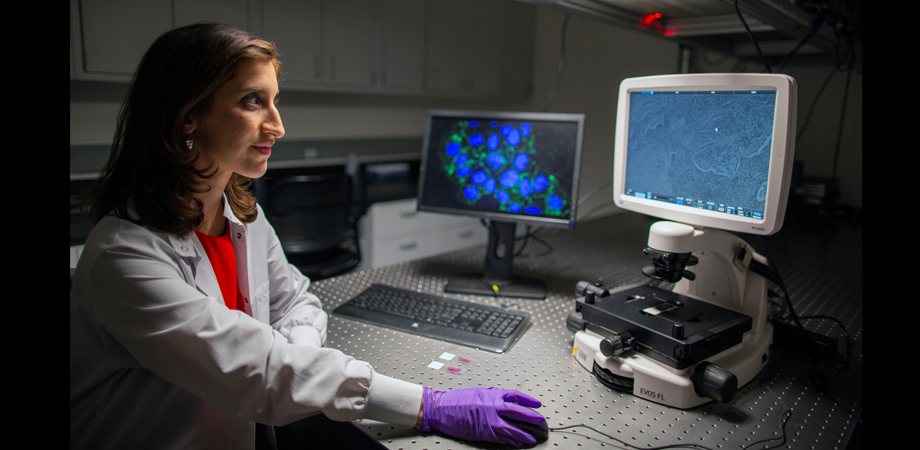
Lisa Poulikakos and her colleague Loza Tadesse study pathological tissue biopsies at Stanford University. Credit: L’Oréal USA For Women in Science
Would you briefly detail some of your lab’s research projects?
Organ fibrosis, the uncontrolled growth and proliferation of fibrous biological tissue, can be related to 1/3 of deaths worldwide, encompassing heart disease, neurodegenerative diseases, and various cancers. However, an incomplete understanding of the role of tissue microstructure in the origin and progression of these diseases hinders the development of effective diagnostic tools and treatments. Existing technologies to image biological tissue microstructure face prohibitive tradeoffs: either their complexity and cost hinders implementation in biomedical and clinical settings, or they lack precision and provide predominantly qualitative information.
My lab's research introduces a new class of metasurfaces which map the microstructural properties of biological tissue onto a quantitative color response on a single, nanostructured optical surface.
What advances and or breakthroughs in the lab are you most excited about?
Structural color is abundant in nature, arising in the brilliant blues of the Morpho butterfly wing or the greens of jeweled beetle shells. We are currently conducting research which draws inspiration from the intricate micro- and nanostructures of these natural photonic systems to develop a new class of nature-inspired colorimetric metasurfaces for biological tissue imaging and diagnostics.
Much of your work involves combining engineering with medicine. What are some of the issues you’ve come across in this work and how have you dealt with them?
UCSD provides the ideal research environment to bridge engineering and medicine. I have been very fortunate to collaborate with world-class researchers at the UCSD School of Medicine, who provide essential medical and clinical insights, as well as renowned colleagues across engineering and the physical sciences with cutting-edge expertise in biomaterials, organoids, and tissue engineering. This highly interdisciplinary research area provides wonderful opportunities for us to broaden our horizons across disciplinary boundaries and develop creative solutions to essential healthcare problems in previously unexplored ways.
What would you like attendees to learn from your upcoming talk on “Colorimetric metasurfaces for next-generation on-chip imaging of tissue microstructure?”
I hope that attendees will learn about the exciting area of nanophotonic metasurfaces for quantitative imaging of biological tissue microstructure as well as the great potential of these novel imaging platforms to be implemented in various clinical and biomedical scenarios. Essential future applications include in-vitro tissue culture imaging, metasurface-aided optical endoscopy, or high-throughput drug screening to address a variety of essential medical challenges.
| Enjoy this article? Get similar news in your inbox |
|


