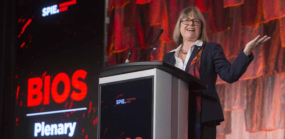
News from SPIE Photonics West 2019
The latest developments in optics and photonics technologies.

Satellite-based optical communications
Researchers gathered at SPIE Photonics West to discuss the latest projects to advance satellite-based optical communication. Optical frequencies, whose data transmission capabilities have long been demonstrated in terrestrial fiber networks, can deliver information much more quickly than conventional radio waves. In recent years, researchers have demonstrated optical links between satellite and ground with the goal of establishing a space-based optical communications network.
Christian Fuchs of the German Aerospace Center (DLR) gave an update on his organization's OSIRIS program, which manages to fit 1550-nanometer optical terminals into a nanosatellite. These small satellites, some the size of a loaf of bread, offer scientists the opportunity to perform more experiments and demonstrations at lower cost. DLR launched the first version of this satellite, the "Flying Laptop," in 2017. The newest version, planned for launch this year, can transmit up to 100 megabits per second.
Several presenters discussed quantum key distribution (QKD), a optics-based cryptographic method that promises high security that some banks and governments are interested in deploying. So far, most demonstrations of QKD have been terrestrially based, where cryptographic keys in the form of photons are transmitted through optical fiber. But the signals die out after about 100 kilometers of transmission, so researchers want to use satellites to transmit the signals. In 2020, DLR plans to launch a nanosatellite-based terminal for quantum key distribution. Stephen Lee of Optocap presented on the development of a compact laser source for satellite-based QKD.
Space-based optical systems have applications outside of communications. Michael Damzen of Imperial College London discussed space-based lasers known as lidar. These instruments are used for remote sensing for monitoring the health of the planet and are used for agriculture management and disaster mitigation, for example. Damzen's team has developed a laser based on alexandrite. One advantage of an alexandrite laser is that its wavelength can be tuned, unlike the neodymium-doped YAG lasers conventionally used in lidar.
But many engineering challenges lie ahead. Space is an unpredictable place-so achieving the necessary robustness is no easy task, said Damzen.
Sophia Chen
Building a Better Clock
Quantum building blocks make inherently good measurement tools. An atom or molecule has one major advantage as a sensor: they are fundamental entities that occur in nature whose properties do not warp over time. For this reason, they make stable sensors that require little to no calibration compared to other types of tools. Now that researchers have invented atomic clocks, quantum thermometers, among other quantum sensors, they are trying to create robust versions that can function outside of the lab-even in space. Researchers presented about these projects at SPIE Photonics West over several days.
On Sunday, 3 February, Jay Lewis of DARPA discussed various efforts to miniaturize quantum technologies. The challenge is to shrink devices that require a table full of optics into a small, energy-efficient package. One success story for the field is the development and commercialization of chip-scale atomic clocks (CSAC), based on which have been commercially available for several years.
In addition, researchers want to make the devices more robust. Researchers have installed the Cold Atom Laboratory on the International Space Station, where it is capable of producing and manipulating delicate quantum states known as Bose-Einstein Condensates. On Monday, Brian Kasch of the Air Force Research Laboratory Lab presented about a compact atomic clock based on a two-photon transition in rubidium that can operate at warmer temperatures than other atomic clocks.
Several presenters discussed the use of laser frequency combs for measurement. Andre Luiten of the University of Adelaide presented on a broadband laser frequency comb that can measure temperature, pressure, and even concentrations of vapor. They use the frequency comb to measure the spectroscopy of groups of molecules. By analyzing the shape of the spectra, they can extract thermodynamic information. Henry Timmers of the National Institute for Standards and Technology presented a new infrared laser frequency comb for studying molecules. The device could be used to study biologically relevant molecules, for example.
Lewis also talked about funding programs for future technologies. The AMBIIENT program at DARPA, for example, is funding the development of quantum magnetometers for biological imaging. These magnetometers could be based on sensors made of atoms in glass cells.
Researchers have demonstrated The most precise clocks today, known as optical lattice clocks, will gain or lose less than a second over the lifetime of the universe, but "good luck keeping it running that long," said Lewis. The next step is making it run longer.
Sophia Chen
Optical Coherence Elastography: Biomechanics of the Eye
The eye is a squishy ball full of springy tissues and membranes. No one knows this better than corneal doctors and scientists, who measure the eye's mechanical properties to guarantee a person's eye health. Still, experts don't understand how changes in eyeball squishiness relate to corneal diseases. They gathered at SPIE Photonics West in February to discuss recent progress in the field, including several studies in which researchers measured the mechanical properties of patients' eyes.
In particular, several speakers focused on a disease known as "keratoconus," an eye disease in which the normally round cornea begins to bulge. The cornea takes on a cone-like shape and thins in the middle, which distorts vision. In severe cases, the patient may require a corneal transplant. The occurrence of the disease is often underestimated, said ophthalmologist J. Bradley Randleman of the University of Southern California. Many people cite that one in 2000 people get the disease, but other studies suggest it could be ten or even 100 times more prevalent.
Currently, ophthalmologists use several techniques to look for keratoconus in patients, which include mapping corneal thickness and measuring the curvature of the eye using techniques such as Scheimpflug imaging. But it's difficult to use these measurements to detect the early stages of the disease. In addition, doctors using two different machines might make completely different treatment recommendations because images can differ so much.
So the field needs to work on developing better mechanical measurements of the eye, said Randleman. In particular, experts don't fully understand the efficacy of a keratoconus treatment known as corneal cross-linking, in which eyedrops and UV light are used to add special bonds between the collagen proteins in the cornea.
Matthew Ford of the Cleveland Clinic presented a study in which his team measured the springiness of patients' eyes before and after a treatment. In the study, they essentially pressed on the patients' eyeball and measured how far it moved given a certain amount of force.
Alfonso Jiménez Villar of Nicolaus Copernicus University developed a model of the cornea so they could better understand the effects of fluid pressure inside the eye. They then compared the model to data collected of air being puffed into subjects' eyes. They found that the model could predict certain effects of the pressure.
Randleman displayed eye maps for two different 20-year-old men, both with corneas with slight asymmetrical bulges and thinning spots. Only one of them developed keratoconus, said Randleman, and it's unclear why. "We need better biomechanical evaluations," he said.
Sophia Chen
Mechanisms of photobiomodulation
In recent years, scientists have begun to investigate a therapy known as photobiomodulation (PBM), which treats ailments by illuminating patients with low levels of light. Patients have used the therapy to accelerate wound recovery, improve their attention, and even regrow hair. But experts don't understand the therapy's underlying biological mechanisms. At SPIE Photonics West, they gathered to discuss the latest studies in the area.
One leading hypothesis on the therapy mechanism is that it increases activity inside mitochondria in cells, said Josh Lalonde of Texas A&M. It could be that the light alters how electrons move within the mitochondria membrane. Lalonde used Raman spectroscopy to investigate a sample of mitochondria before and after illumination. In particular, he studied PBM's effects on a particular protein inside the mitochondria called cytochrome c.
Ali Shuaib of Kuwait University discussed how PBM studies do not report standardized metrics, which makes it difficult to compare different studies. For example, researchers inconsistently report fluence, which is the amount of energy that light deposits at an injury site. Shuaib's team simulated a rat with a spinal cord injury and modeled how a low-energy laser beam propagated through the rat's body. They created and compared the results of more than 900 simulations, in which they varied the laser beam diameter and the orientation of the rat's body. They plan to simulate this in a human model next.
Daqing Piao of Oklahoma State University also conducted a study on how light propagates during the therapy-but on actual animal tissue. His team measured how light penetrated the skin of cadaver dogs they'd gotten from an animal shelter. They illuminated several areas along the cadaver's spine and found, for example, that only 12 percent of light would transmit through the skin. They also found that applying the light source while making contact with the skin increases the transmission by 68 percent. They plan to run the same studies using a horse cadaver next.
Justin Rigby of the University of Utah conducted a study on the efficacy of PBM for alleviating muscle fatigue. Athletes with muscle fatigue are more likely to get injured. They performed a controlled study on 34 college-age participants by having them flex their arm before and after illumination with red and blue light. In this preliminary work, Rigby said that PBM shows promise for lessening muscle fatigue, but they will have to conduct more studies.
Researchers are also studying whether PMB might be helpful against cancer. Sergei Sokolovski of University of Aston presented on a study in which low levels of infrared light were beamed onto colon cancer cells. They could tell that the cells died at different rates depending on the light dose, and that the light disrupted mitochondrial functioning. By understanding better how PMB works and optimizing the various techniques involved, experts hope to apply the therapy more effectively to treat disease.
Sophia Chen
Photonics in Dermatology and Plastic Surgery
On Saturday, researchers gathered at SPIE Photonics West 2019 to discuss methods to monitor and improve skin health using light. The techniques use a range of wavelengths and target various medical applications, including wound recovery and better sunscreens.
Kapil Dev of the Singapore Bioimaging Consortium, for example, is using a technique known as raster scanning optoacoustic mesoscopy (RSOM) for imaging inflammatory skin conditions. When applied to skin, an RSOM instrument delivers nanosecond-long pulses of light to produce ultrasound waves in a broad range of frequencies. The advantage of the technique is that it can image a considerable depth-several millimeters beneath the skin-while also maintaining good image quality.
These 3D RSOM images, explained Dev, contain quantifiable information about the skin that doctors can use to treat patients. Dev's team has been using RSOM to study eczema. Conventionally, doctors determine the severity of eczema by visually examining the lesions and asking the patient about their levels of discomfort. This is a relatively subjective process. RSOM images provide doctors with more objective metrics. For example, they have found that a patch of skin with a thicker epidermis, higher blood volume, and denser capillary networks in the area corresponds to a more severe case of eczema.
Hanna Jonasson of Linköping University in Sweden is studying the optical scattering and absorption properties of human skin. To do this, her team has created a model of skin and how various layers in the skin interact with light. Then, they collected optical data from the skin of nearly 1,800 people, roughly half women and half men. By interpreting the data within their model, they found that women's skin, on average, scatter light differently than men's.
This data is part of an ongoing program in Sweden called the Swedish Cardiopulmonary Bioimage Study (SCAPIS) dedicated to preventing cardiovascular and pulmonary disease in Sweden. SCAPIS will produce a large open-source data set whose statistics can serve as a reference by researchers around the world, said Jonasson.
Ying Wang, a doctor and researcher at Massachusetts General Hospital, studies how wounds heal. "My patients often ask me, ‘Do you think my wound has healed?'" Wang said in the presentation. "Most of the time I can't answer that question." That's because doctors still largely rely on their own eyes-"gross visual assessment," as Wang calls it-to determine how quickly a wound will heal.
To improve these assessments, her team has developed a fluorescence imaging technique to monitor a wound's progression in tissue culture and on rat tails. As a wound heals, skin cells that contain the amino acid tryptophan and the protein collagen begin to proliferate on the area. Under ultraviolet light, these compounds fluoresce. Wang's team applied UV to these tissues and found that as the wound healed, it would fluoresce more intensely up to a certain point. They think that these UV fluorescence images could be used to evaluate the severity of wounds and their healing time and are pursuing clinical studies.
Steven J. Davis, a physicist from Physical Sciences Inc. based in Massachusetts, is developing an instrument for measuring so-called singlet oxygen, a molecule produced when ultraviolet light ranging from 340 to 400 nanometers, known as UVA, interact with human skin. Exposure to UVA, which penetrates skin more deeply than shorter-wavelength UVB, increases skin cancer risk. However, most commercial sunscreens do not protect against UVA.
Davis thinks his instrument could help evaluate the effectiveness of sunscreens against UVA. To test their instrument, his team recruited 12 people through the University of California, Davis. Each person placed a forearm inside a chamber that irradiated it with UVA, and Davis showed they could detect levels of singlet oxygen produced.
During the question-and-answer after the talks, several audience members expressed interest in using these techniques in their own laboratories. Although researchers demonstrated the methods on specific medical applications, several could be broadly useful in a clinical setting: one audience member, for example, pointed out that Dev's instrument could be extended to monitor skin conditions other than eczema.
Sophia Chen
| Enjoy this article? Get similar news in your inbox |
|


