Through a Metalens, Lightly
There is a clinical need for tools that can accurately visualize lesions in internal organs, making diagnosis more accurate, Hamid Pahlevaninezhad told the audience at SPIE Photonics West in February. The Harvard Medical School research fellow noted that diagnosis of diseases in internal organs rely mainly on the histological evaluations of the samples collected from affected regions, and poor diagnostics are often due to the inability of current instruments to locate and sample lesions.
"Optical coherence tomography, or OCT, is one of the promising techniques to address this need, said Pahlevaninezhad. "It provides about 10-micron axial resolution, about 20 to 30-micron lateral resolution, and about 1.5-millimeter penetration depth into the tissue."
As a part of the Optical Coherence Tomography and Coherence Domain Optical Methods in Biomedicine session, Pahlevaninezhad discussed a nano-optic endoscope for high-resolution endoscopic OCT, a project he's been working on at Melissa Suter's lab at Massachusetts General Hospital (MGH). Suter is an assistant professor at MGH and Harvard Medical School.
Endoscopic high-resolution optical imaging can enable several applications in diagnosing disease in internal organs. However, difficulties associated with optical aberrations and the trade-off between transverse resolution and depth-of-focus significantly limits the scope of applications.
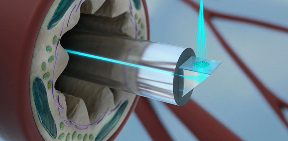
The work from Suter's lab presents a new class of optical imaging catheters -- nano-optic endoscopes -- that address these difficulties. A new dimension to the lab's work has been added by partnering with Federico Capasso's lab in Harvard's John A. Paulson School of Engineering and Applied Sciences (SEAS). Capasso's group at SEAS have been developing metalenses to achieve wavefront shaping of light using optical elements with thicknesses on the order of the wavelength. This miniaturization could to lead to compact, nanoscale optical devices with applications in cameras, lighting, displays, and wearable optics.

The nano-optic endoscope design: (A) a single-mode fiber is housed in a drive cable on a ferrule. A prism reflects light toward the metalens. (B) The fabricated nano-optic endoscope. (C) A metalens building block. (D) An SEM image of part of the fabricated metalens.
The method Suter and Capasso's teams are working on incorporates such a metalens into the design of an OCT catheter to achieve near diffraction-limited imaging at extended depth-of-focus by negating non-chromatic aberrations and chromatic dispersion engineering. The design versatility of the nano-optic endoscope can significantly elevate endoscopic imaging capabilities.
"Essentially, we can take these big, bulk optics with all the things that they do and design them into a single, thin wafer," said Suter in a post-presentation. "This allows us to precisely control things that we can't necessarily do with these big optics -- it just opens up the design possibilities and other things we can do with catheters."
Location, location, location
According to Suter, one key to her project's success is the location of her lab -- two floors above the pathology section at MGH. "We have excellent collaborators and co-investigators there," she said. Following MGH-approved protocols, Dr. Lida Hariri, an assistant professor in pathology, can bring tissue samples to image just prior to submitting them for histopathology processing. "A board-certified pathologist is in control of the tissue at all times," Suter added. "We're lucky to have this access to fresh samples."
In addition, MGH has several large-animal facilities on the main campus, which makes it easier for Suter's lab to do more translational work than offsite labs that don't have close access to pathology and surgery. "I think that's really made a huge difference for us," she said.
To check the image quality of the nano-optic endoscope and the effect of removing aberrations, the team used a grape, as fruit flesh has fairly uniform cellular structures. Features are defined in lateral and axial directions.
Yet another strength has been the team's ability to combine engineering and medical technology. With his background in electrical engineering and physics, Pahlevaninezhad has found a few challenges working in a medical imaging lab: in the engineering world, the speed of translation can be very fast, with changes often occurring on a daily basis. But in the medical field, he noted, it can be difficult to change things. In a medical lab, a new method or tool must be thoroughly tested and proven to be much better than what's already available, and that takes time.
"But the most amazing thing to me is when you design something, and it works the way you want it to, and it's something that really matters. For example, we calculated the resolution, and when we measured, it was within less than 1 micron of what we expected. So, calculations translate to what we build, and that's kind of satisfying for me."
Getting better all the time
In a 2013 interview with SPIE TV, Suter discussed next-generation OCT for complex imaging of the lung. When asked what advances she has seen in the technology since then, she emphasized contrast resolution, "particularly the resolution that we've been able to push, specific to applications in pulmonary," she said. "One of the big things was the resolution for lung cancer work that Hamid presented -- that's really going to allow us the see these pathologic features. The other is the contrast -- we've done a lot of work in polarization sensitive imaging, specifically tailored for our asthma work, and that's really changed what we can assess in smooth muscle such as contractile force that we previously couldn't see with our OCT or OFDI. Those have been the two biggest game changers recently for us."
From Suter's point of view, the work done so far is just the beginning. Metalenses can be designed to do several things, such as precisely controlling light in ways that are normally difficult with other optics. "Not only is it essentially a small wafer that we're placing on the end of the catheter, but the opportunity to precisely control the light is huge and that's just going to open the door to multiple different imaging techniques. We use OCT because that's what we work with, but it could be used for visible to near infrared light as well. The potential is endless and what we've done so far is just the tip of the iceberg."
Related SPIE content:
Hamid Pahlevaninezhad: High-resolution endoscopic optical coherence tomography of airways
Federico Capasso: Flat optics: from high-performance metalenses to structured light
Federico Capasso: Metasurface flat optics: Unifying semiconductor manufacturing and lens making
| Enjoy this article? Get similar news in your inbox |
|


