Imaging Blood Vessels at the Speed of Sound
A research group at Caltech uses photoacoustic computed tomography to visualize blood flow in extremities.
Photoacoustic imaging is a kind of oddball imaging modality. The basic idea is, like all good ideas, simple. When a pulse of light is partially absorbed by matter, the matter rapidly expands. The expansion generates a pulse of sound waves, which are detected by a set of ultrasound transducers, or microphones. Then, the location of the absorbing material is calculated by triangulation. With a bit of computation, a 3D image of a structure can be obtained.
This combination of excitation via light and detection via sound makes photoacoustic imaging very attractive for imaging inside the body. Contrast between different tissues can, to some extent, be controlled by choosing the right wavelength of light. Light has a short wavelength, compared to sound, so the spatial resolution is very high.
Using light to generate sound also creates a relatively clean signal. Acoustic waves are not so strongly absorbed or scattered as light waves, so the received acoustic signal is comparatively simple to interpret.
The downside is that photoacoustic imaging is agonizingly slow, and may require the subject to hold absolutely still for a good 30 minutes. However, new research from Lihong Wang's laboratory at CalTech suggests that improvements in photoacoustic imaging speed are imminent.
A snail's pace
To understand why photoacoustic imaging takes so long, we need to delve into the physics a little bit. If you hold your hand over a flashlight, you will see a bright red glow. The light from the flashlight manages to pass right through the tissue and exit the skin. But, you can't see the flashlight itself, because the light does not take a direct path through the tissue to your eye. Every cell membrane, vesicle, and other sundry biological bit redirects the light, scattering it into a diffuse glow.
In conventional photoacoustic imaging, a laser emits ten pulses per second to illuminate a small patch of skin. The emitted light fills a large tissue volume, diluting the energy density of the beam. The corresponding acoustic signal is small, so a large number of light pulses may be required to obtain a sufficiently clear signal.
To get high-quality images, you need a lot of microphones. The data from each microphone then needs to be sent to a computer fast enough to generate an image.
The laser and microphone array may also be scanned to image larger tissue sections, further increasing the time it takes to record an image.
The speed of sound
To solve these problems, Wang's research group scaled up the whole imaging system. The laser they used has sufficient pulse energy to illuminate a large area of tissue from two sides simultaneously. The microphone array was expanded to an astounding 512 microphones, placed in a ring around the illuminated tissue. And in a fit of enthusiastic engineering, the 512 microphones were sampled simultaneously using a custom data acquisition system.
That means that a single slice image takes only 100 microseconds to acquire. Large 3D images are obtained by scanning the microphone array. Taken together, a full image is generated in 5 to 15 seconds. That in itself is a huge improvement. But, the single shot speed is also a big gain here.
The point is that each shot produces a crisp image, and each image overlaps somewhat with its neighbors. So, even if the patient moves during imaging, the sharpness allows the images to be stitched together, creating a beautifully clear 3D image.
Revealing damaged blood vessels
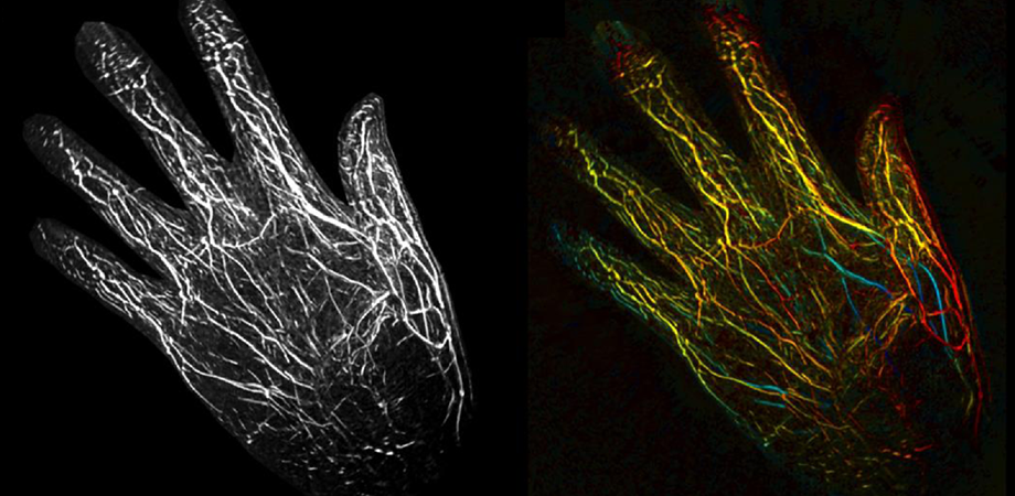
In this work, researchers were able to image blood vessels to a depth of nearly 2 cm. And, because light around 700-800 nm wavelengths is known to be able to image much deeper into the body, future work may examine customized light sources that will allow fast photoacoustic imaging to be used on internal organs and more deeply buried blood vessels.
However, the 2 cm imaging depth is quite sufficient for imaging blood vessels and blood flow in hands, fingers, and toes, which are fairly thin appendages. The sharp images of this system, combined with the delay between slices, may help doctors to diagnose vascular disease. The researchers showed that normal blood vessel motion is clearly visible in the photoacoustic images due to the high optical absorption of hemoglobin. Blood vessels move as the heart pumps, but that motion changes when the vessels are diseased. With improvements to imaging speed and increased applications, photoacoustic imaging is becoming an audible contender for widespread clinical adoption.
Read the article on the SPIE Digital Library: J. Biomed. Opt. 24(2), 026501 (2019) https://doi.org/10.1117/1.JBO.24.2.026003
Chris Lee is a physicist and writer living and working in Eindoven, the Netherlands.
| Enjoy this article? Get similar news in your inbox |
|


